THOMALE GUIDE
FEATURES
- controlled placement of the ventricular catheter
- low technical effort
- together with the THOMALE GUIDE APP for the iPhone and iPad a simple navigation and guidance aid
- especially for the placement of ventricular catheters in narrow ventricles
Instrument
One risk factor for a possible disruption of CSF drainage through the ventricular catheter is incorrect placement. In the past, catheters were usually placed freehand. The literature shows that in more than 40% of cases* the ventricular catheter is incorrectly positioned. Therefore, in recent years, aids such as ultrasound or neuronavigation have been increasingly used to better control the implantation of ventricular catheters. However, this requires a high technical effort fü̈r a relatively minor surgical procedure. The THOMALE GUIDE was developed to make the placement of the ventricular catheter more precise and less complicated with little technical effort.
* 2008, Huyette, J. et al. „Accuracy of the freehand pass technique for ventriculostomy catheter placement: retrospective assessment using computed tomography scans“, J Neurosurg 108(1): p.88-91
The THOMALE GUIDE consists of four individual parts: Catheter guide (1), locking nut (2), glider (3) and rails with scale and base ring (4). It was developed for the MIETHKE ventricular catheter with an inner diameter of 2.6 mm. Two further catheter guides with inner diameters of 3.1 mm and 3.6 mm are also included.

Functionality
The THOMAL GUIDE allows guided puncture of the ventricle with a ventricular catheter via a frontal (precoronary) borehole. The boreholes start already 3 mm from the tip and end after 14 mm. This prevents drainage from the ventricles into the brain when implanting in very narrow ventricles.
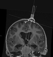
For a controlled placement of the ventricular catheter, the THOMAL GUIDE is placed directly on the skull bone with three feet and with the line markings parallel to the centerline above the drill hole to ensure a right angle of insertion in the sagittal plane. In the coronal plane, an individual angle can be determined using an angle adjustment on the THOMAL GUIDE. The rotation of the catheter guide, which is guided over the rails, is done via a pivot point located very close above the borehole.
In order to measure the insertion angle, it is necessary in principle to determine the desired angle of the ventricular catheter to the tangent in the coronal plane above the borehole.
There are two methods for this:
1. The most precise method for determining the individual insertion angle of the ventricular catheter requires the reading of a volume or thin-film data set into a planning software. In this software the optimal trajectory for the ventricular catheter can be defined. The coordinates of the entry point, e.g. to the nasion and the centerline, can be measured. At the entry point, the tangent would have to be drawn in the projected coronal plane and the angle of the trajectory to the tangent measured. Now the data of the entry point can be transferred to the patient and intraoperatively the angle can be applied on the THOMAL GUIDE for the insertion of the catheter. This method is especially recommended for very narrow ventricle systems. The accuracy of determining the angle depends on the image used.


A coronary sectional image should be used to facilitate individual measurement of the angle of entry. If possible, this should be at the level of the anterior horn in front of the Foramen Monroi. The entry point, the projected target point and the projected trajectory should be marked on the foramen monroi. To mark the projected target point, the distance to the midline and the depth of the ventricle should be taken into account. Now the tangent can be drawn. The easiest way to do this is to set two points at equal distances medially and laterally from the entry point. The connection of these two points corresponds to the tangent. Accordingly, the angle between the trajectory and the tangent can then be determined and set on the THOMAL GUIDE.


For both methods the THOMALE GUIDE App for iPhone and iPad will be used. It allows - e.g. via an image import - a simple determination of the angle of the trajectory to the tangent.
DO YOU HAVE ANY QUESTIONS ABOUT THE PRODUCT?
WE ARE THERE FOR YOU

OUR PARTNERSHIP
WITH B. BRAUN
B. Braun and MIETHKE - Together for a better life with hydrocephalus
We have a long and intensive partnership with B. Braun in the field of neurosurgery. We are driven by a common vision: to improve the lives of hydrocephalus patients around the world with innovative solutions.
Our partnership is an exciting combination of B. Braun's many years of expertise as one of the world's leading medical device and pharmaceutical companies and our agility as an innovative company and technology leader in gravitation-based shunt technology.
Our Strong Partner in Neurosurgery:


